 FUNCTIONAL MAGNETIC RESONANCE IMAGING (fMRI)
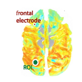
Functional magnetic resonance imaging (fMRI) is among the youngest techniques in the neuroscientific arsenal. fMRI is used to measure local changes in the brain's blood supply caused by focal neural activation.
One major strength of fMRI lies in its global coverage of slow varying activity, which renders it an ideal counterpart to the fast, but spatialy limited methods used in neurophysiology.
Our lab specializes in the combination of electrical measurements of local neuronal activity and whole-brain assessments using fMRI. The results of these studies affect both our knowledge of the neural events causing the fMRI signal as well as our understanding of the interplay between large scale neural networks and localized synaptic processing.
REPRESENTATIVE PUBLICATIONS:
Leopold, D.A. & Maier, A. (2011)
Ongoing physiological processes in the cerebral cortex.
Scholvinck, M.L., Maier, A., Ye, F.Q., Duyn, J.H., Leopold, D.A. (2010)
Neural basis of global resting-state fMRI activity.
Proc. Natl. Acad. Sci. USA 107(22):10238-43
Maier, A., Wilke, M., Aura, C., Zhu, C., Ye, F.Q. & Leopold, D.A. (2008)
Divergence of fMRI and neural signals in V1 during perceptual suppression in the awake monkey.
Nat. Neurosci. 11(10):1193-1200
[see also: DISPATCH: Blake, R. & Braun, J.. (2009). Curr. Biol. 19(10):R30-32]
